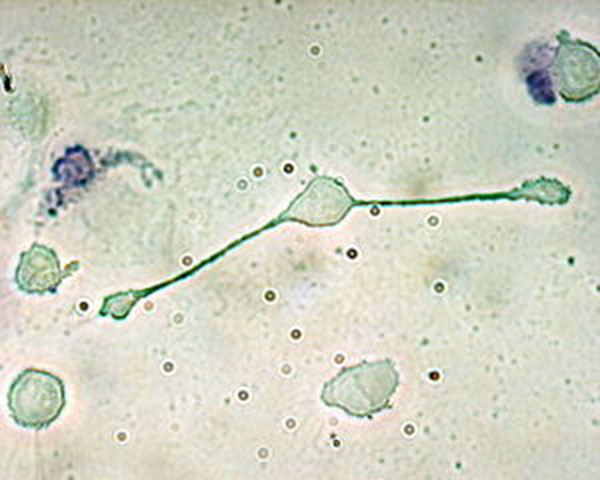
Macrophages are a class of malleable cells that can be polarized to Type 1 macrophages (M1) and Type 2 macrophages (M2) under different microenvironments, and are different in wound repair The stage plays a central role.

Macrophage polarization is based on the induction of different cytokines by Th1 and Th2 cells. Macrophages are activated to proinflammatory (M1, also known as the classically activated macrophages), stimulated by cytokines secreted by Th1 cells [such as interferon-γ and tumor necrosis factor-α] And classically activated macrophages); whereas Th2 cytokines such as IL-4, IL-10 and IL-13 then polarize macrophages into anti-inflammatory forms (M2, also known as selectively activated macrophages) activated macrophages). In general, M1 macrophages have the phenotype of IL-12hiIL-23hiIL-10low, producing effector molecules such as reactive oxygen species and active nitrogen mediators, as well as various inflammatory cytokines such as IL-1β, TNF-α and IL- 6, etc.) have the function of killing microorganisms and play an important role in the early stage of trauma. While type M2 macrophages are characterized by IL-12 low IL-23 low IL-10 hi, high expression of the scavenger receptor, mannose receptor and galactose-type receptor, Secretion of anti-inflammatory factors (such as IL-10, etc.), a variety of extracellular matrix proteins [such as fibronectin, transforming growth factor-β inducible gene-H3 and so on] and a variety of proangiogenic factors [ Such as TGF-β1, platelet-derived growth factor subunit-B, hepatocyte growth factor (HGF), and insulin-like growth factors (IGF-1), promote cell proliferation, collagen deposition and blood vessels Generate play a leading role.
M2 macrophages are an indispensable subset of macrophages in wound healing and can be further differentiated into three subtypes under different microenvironmental stimuli: (1) M2a type, which can be activated by IL-4 or IL -13 stimulation, with the promotion of fibrosis; (2) M2b type, Toll-like receptor (or IL-1 receptor) combined with immune complexes can induce this type; (3) M2c type, IL- -β or glucocorticoids can stimulate the formation of M2c not only has immunosuppressive function can also promote the formation of new blood vessels.
Researchers have come up with different hypotheses about the origin of traumatic local M2 macrophages. The first view is that specific monocyte subpopulations migrate and differentiate into specific subtypes of macrophages. For example, Ly6Chi monocytes differentiate into M1 macrophages and Ly6Clow monocytes differentiate into M2 macrophages. However, some researchers have observed that Ly6Chi monocytes can also differentiate into M2 macrophages and Ly6Clow monocytes can be transformed into M1 macrophages, and M1 and M2 macrophages can transform into each other. The second view is that circulating monocytes in different parts of the trauma at different times to local differentiation can be divided into different subtypes, and that this is the inflammation of the micro-environment at the decision. The third view is that trauma local M2 macrophages are transformed from M1 macrophages under certain microenvironment conditions. Researchers have succeeded in using microRNA-21 to convert proinflammatory macrophages to anti-inflammatory forms, and believe this successful transition is due to the modification of intracellular non-coding small RNA microRNA-21 or the modification of microRNA-21 Target protein tumor suppressor gene homologous phosphatase – tensin homolog deleted on chromosome ten (PTEN) and programmed cell death 4 (programmed cell death 4, PDCD4) levels caused by changes.
Leave a Reply